Case of the Fortnight #1
Clinical history:
68-year-old male patient with rapidly growing swelling of 4 months duration. On the dorsal aspect of the forearm. O/e fairly well-circumscribed flesh-colored nodule., 2 x 1.8 cm. No history of other tumors in the body.
CASE OF THE FORTNIGHT Dr Gowripriya Consultant pathologist Dr Rela institute and medical center
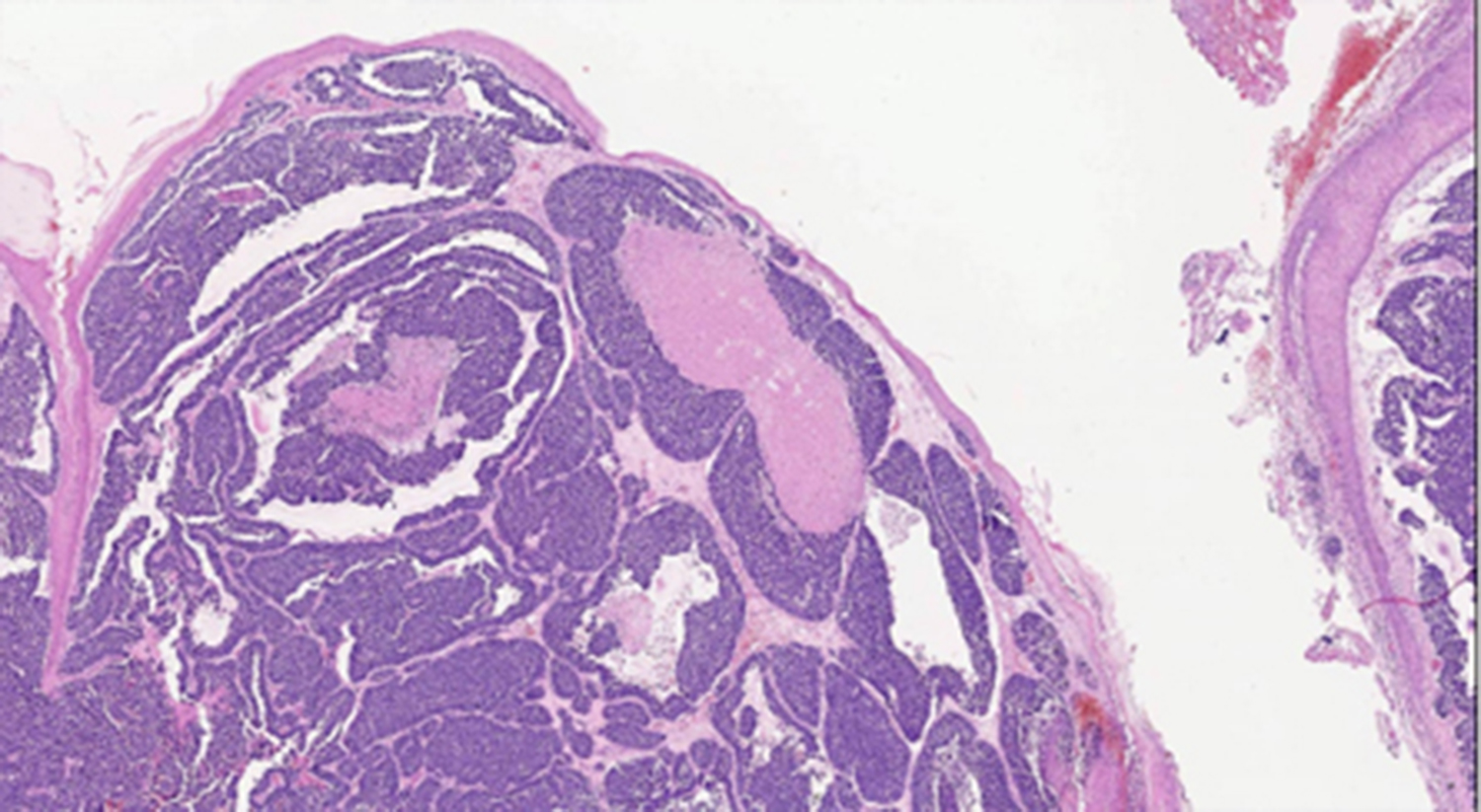
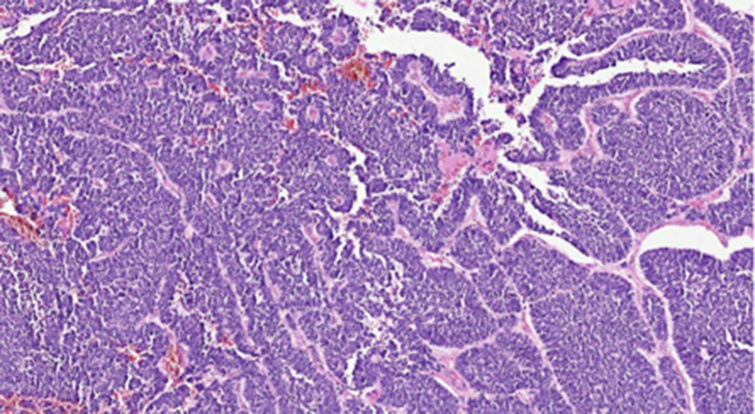
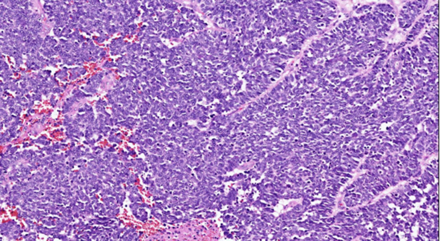
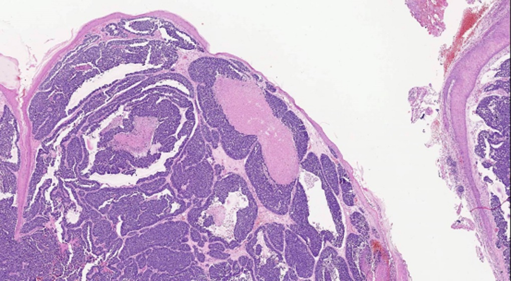
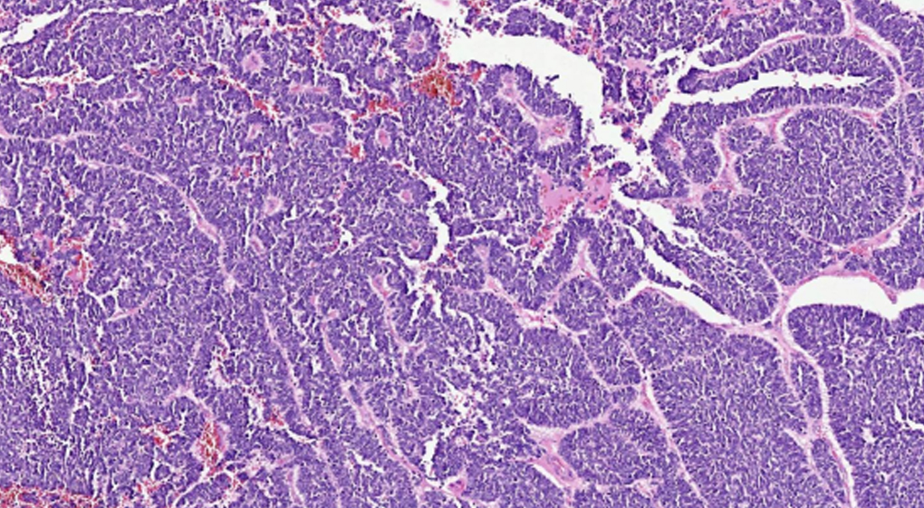
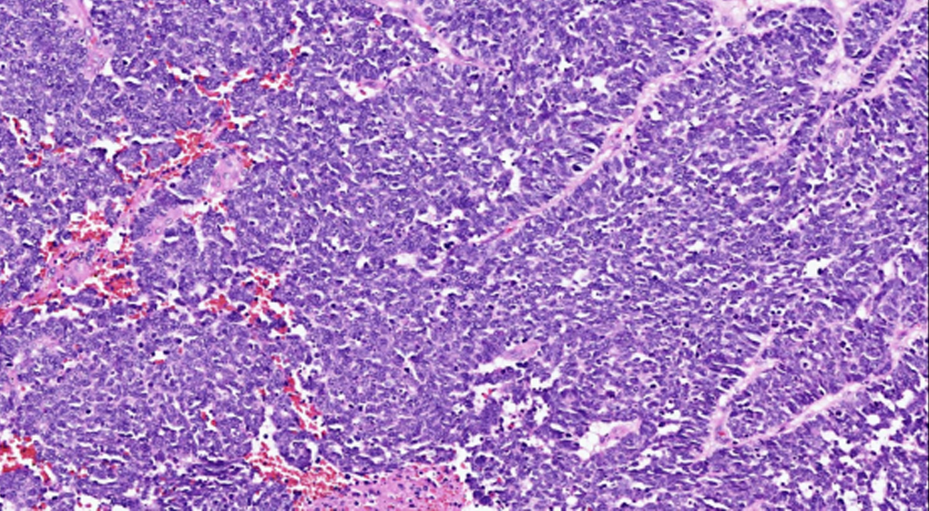
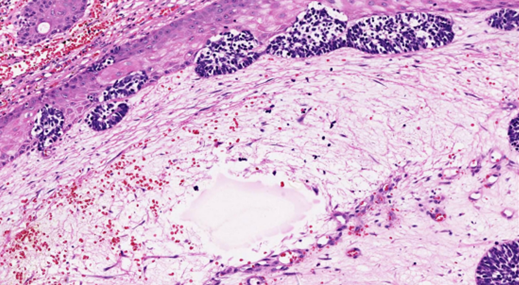
MERKEL CELL CARCINOMA
- 68 YEAR OLD MALE PATIENT WITH RAPIDLY GROWING SWELLING OF 4 MONTHS DURATION.
- ON THE DORSAL ASPECT OF FOREARM.
- O/E FAIRLY WELL CIRCUMSCRIBED FLESH COLOURED NODULE., 2 X 1.8 CM.
- NO HISTORY OF OTHER TUMOURS IN THE BODY.
- 68 YEAR OLD MALE PATIENT WITH RAPIDLY GROWING SWELLING OF 4 MONTHS DURATION.
- ON THE DORSAL ASPECT OF FOREARM.
- O/E FAIRLY WELL CIRCUMSCRIBED FLESH COLOURED NODULE., 2 X 1.8 CM.
- NO HISTORY OF OTHER TUMOURS IN THE BODY.
LEARNING POINTS:
- 1)A rare, aggressive, primary cutaneous neuroendocrine carcinoma.
- 2)Elderly males, sun exposed skin of head and neck/ extremities/ trunk
- 3)Immunosuppression and advanced age are risk factors.v
- 4)Cell of origin- unknown, possible precursors include Pro-B/ Pre-B lymphocytes, fibroblasts, dermal mesenchymal stem cells and epithelial cells.
- 5) Clinically, rapidly growing violaceous/ flesh coloured nodules.
- 6)Microscopy: Small blue round cell tumour in the dermis and/ or subcutis.
-
- Cells with high nuclear: cytoplasmic ratio, salt and pepper chromatin.
- Sheets/ nests/ occasional trabeculae/ pseudo-rosettes
- Mitoses/ apoptosis/ necrosis.
- Squamous differentiation maybe seen.
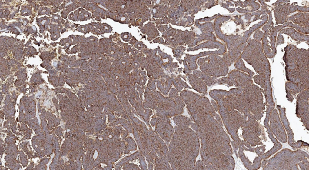
- 7)Immunohistochemistry:
-
- Neuroendocrine markers
- CK 20- paranuclear dot- like positivity
- Neurofilament
- SAT B2
- Often , Merkel Cell Polyoma Virus (see below)
- 8)TWO MAJOR SUBSETS BASED ON VIRAL STATUS:
- 9)Treatment: Excision, lymph node resections, Immune-checkpoint-inhibitors.
- 10)Data for inclusion in pathology reports:
-
- Maximum dimension of tumor (gross or microscopic)
- Tumor confined to dermis/subcutis: yes/no
- Involvement of underlying muscle, fascia, bone or cartilage: yes/no
- Lymphovascular involvement: yes/no
- Distance from surgical margin
- Local microscopic “satellite” deposits of tumor: yes/no
- Distance between main tumor and deep surgical margin
- Morphology (pure neuroendocrine or combined): pure or combined
- Merkel cell polyomavirus status: positive or negative
- Immunohistchemical profile
- P63 expression: positive or negative
| Feature | MCPyV-positive | MCPyV-negative (UV exposure related) |
|---|---|---|
| Incidence | More common | Less common |
| Morphology | Pure | Pure/ combined |
| Prognosis | Better | Worse |
| Tumor infiltrating lymphocytes |
Brisk | Few |
| IHC | Rb pos (+++) P53 pos (+) P63 pos (+) |
Rb pos (+) P53 pos (+++) P63 pos (+++) |
| Genetics | Tumour mutational burden | Tumour mutational burden |
| low No Recurrent RB1/ P53 mutations No UV mutation signature | high Recurrent RB1/ P53 mutations UV mutation signature |
REFERENCES:
1)Noreen M. Walsh, Lorenzo Cerroni; Merkel cell carcinoma: a review, Journal of cutaneous pathology https://doi.org/10.1111/cup.13910
2)WHO classifcation of skin tumours, 4th ed.
